Large Travel Inspection Microscope
Infrared observation can be conducted with the IR objective lenses, which enable the operators to nondestructively inspect the inside of IC chips packed and mounted on a PCB, utilizing the characteristics of silicon that transmit infrared light. 5X to 100X IR objectives are available with chromatic aberration correction from visible light wavelengths through the near infrared. Especially with an objective lens of 20X or more, the aberration caused by the silicon layer covering the observation object can be corrected by the correction collar to obtain a clear image.

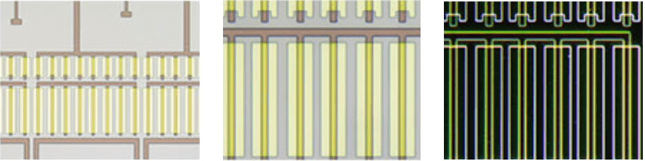
Darkfield is used for detecting minute scratches or flaws on a sample or inspecting samples with mirrored surfaces, such as wafers. MIX illumination enables users to view both patterns and colors.

Photoresist residue on a semiconductor wafer
Fluorescence is used for samples that emit light when illuminated with a specially designed filter cube. This is used to detect contamination and photoresist residue. MIX illumination enables the observation of both the photoresist residue and IC pattern.

A hard disk
Differential interference contrast (DIC) is used to help view samples with minute height differences. It is ideal for inspections of samples having very minute height differences such as magnetic heads, hard-disk media, and polished wafers.

Film
Polarized light is used to reveal a material’s texture and the condition of crystals. It is suitable for inspections of wafer and LCD structures.

An LCD color filter
This observation technique is suitable for transparent samples such as LCDs, plastics, and glass materials. MIX illumination enables the observation of both the filter color and circuit pattern.

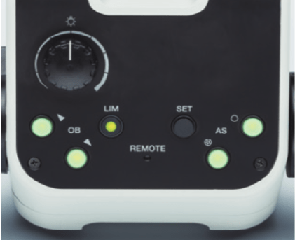
In normal microscopes, users need to adjust the light intensity and aperture for every observation.
The image gets too bright or dark when changing magnification or observation method.
User need to adjust to optimum brightness when changing from Brightfield to Darkfield

Light intensity is automatically adjusted when changing from Brightfield to Darkfield
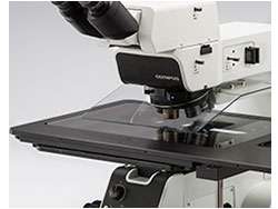
8" Travel microscope ( Stroke: 210mm x 210mm )
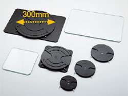
Chose from the wide range of accessories from 6&8 inch wafer holder or stage suitable for panel inspection.
- 3 & 4 inch wafer holder
- 4 & 5 inch wafer holder
- 5 & 6 inch wafer holder
- 6 & 8 inch stage
- 6 & 8 inch rotatable wafer holder
- Flat reflected stage plate
- Glass stage plate

12" Travel microscope (Stroke: 356mm x 305mm)
Chose from the wide range of accessories from 6&8, 8&12 inch wafer holder. The modular design makes it easy to customize the microscope for your specific requirements
- 6 & 8 inch stage
- 6 & 8 inch rotatable wafer holder
- 8 & 12 inch stage
- 8 & 12 inch rotatable wafer holder
- 6" x 6" mask holder
- Glass stage plate

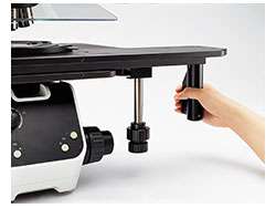
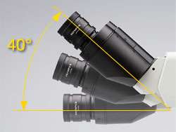
The tilting observation tube’s extensive range enables operators to sit at the microscope in a comfortable posture.
|
|
MX63 |
MX63L |
|
|
Optical system |
UIS2 optical system (infinity-corrected system) |
||
|
Microscope frame |
Reflected light illumination |
White LED(with Light Intensity Manager) 12 V 100 W halogen lamp, 100W mercury lamp, light guide source |
|
|
Transmitted light illumination |
Transmitted light illumination unit: MX-TILLA or MX-TILLB is required. |
||
|
Focus |
Stroke: 32 mm |
||
|
Maximum load weight (including stage and holder) |
8 kg |
15 kg |
|
|
Observation tube |
Wide-field (FN 22 mm) |
Erect and trinocular: U-ETR4 |
|
|
Super-wide-field (FN 26.5 mm) |
Erect, tilting and trinocular: MX-SWETTR (optical path switchover 100% (eyepiece) : 0 (camera) or 0 : 100%) |
||
|
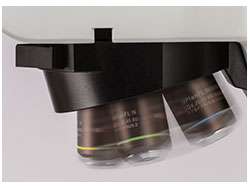
Motorized nosepiece |
Brightfield Brightfield and darkfield |
||
|
Stage (X × Y) |
Coaxial right handle with built-in clutch drive: MX-SIC8R Coaxial right handle with built-in clutch drive: MX-SIC6R2 |
Coaxial right handle with built-in clutch drive: MX-SIC1412R2 |
|
|
Weight |
Approx. 35.6kg(Microscope frame 26kg) |
Approx. 44kg(Microscope frame 28.5kg) |
|
Specification are subject to change without notice or obligation on the part of the manufacturer


